Sheep Eye Dissection
We'll dissect a sheep eye to learn about the different parts of the eye and how it mimics a human eye.Interested in getting your very own BioBox Labs subscri.

Sheep eye Diagram Quizlet
sheep eye dissection virtual practical exam - practice quiz for anatomy - YouTube © 2023 Google LLC Cornea: The outer, transparent structure at the front of the eye that covers the iris,.
PPT Sheep’s Eye Dissection Inside & Out PowerPoint Presentation ID
Close up of the gloved hands of ansnatomy student dissecting the eyeball of a sheep using scissors with samples of the surrounding muscle tissue from the socket lying alongside on the dissecting tray Lama looking straight to the camera
Sheep Eye Dissection
Start studying sheep eye layers. Learn vocabulary, terms, and more with flashcards, games, and other study tools. Fresh features from the #1 AI-enhanced learning platform.
Sheep Eye Dissection
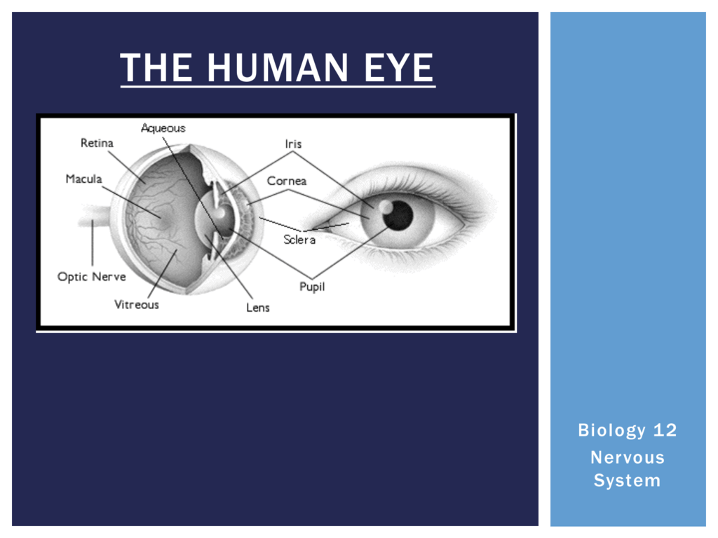
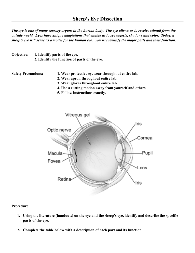
fluid in the eye, found between the cornea and the lens. Vitreous Humor. jellylike substance found behind the lens in the posterior cavity of the eye that maintains its shape. Start studying Lab #12: Sheep Eye Dissection. Learn vocabulary, terms, and more with flashcards, games, and other study tools.
Observations Sheep Eye Dissection Lab
Place the sheep's eye on it for inspection. 2. Use a pencil to sketch a side-view external diagram of the eyeball in Observation #1. Make sure to label the sclera, cornea, optic nerve, and muscles. 3. Cut the fat and muscles off the eyeball so that you can see the sclera. 4. Place your eye specimen in the dissection pan.
16 Sheep Eye Dissection 17 Sheep Eye Dissection Lab
Intro Prof. Wilson sheep eye dissection. The best sheep eye dissection mike watson 413 subscribers Subscribe Subscribed 197K views 11 years ago Prof. Wilson Prof. Sally Wilson dissects a.

Dissection of a sheep eye Diagram Quizlet
Step 1: Wash the sheep eye in running water to remove the preservative fluid. Dry the eye with paper toweling. Examine the front of the eye and locate the eye-lid, cornea, sclera (white of the eye) and fatty tissue. Examine the back of the eye and find extrinsic muscle bundles, fatty tissue and the optic nerve.

Sheep Eye Dissection by Wetmore Gt
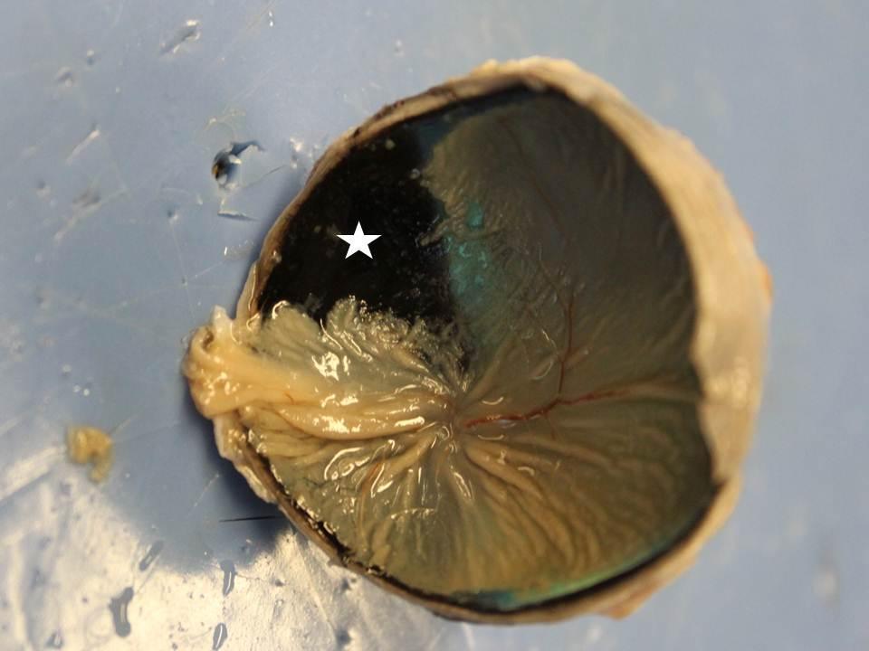
Move the retina to see the dark, metallic-looking tissue at the back of the eye. This is the choroid. The portion that appears iridescent blue and green with shades of yellow is called the tapetum. Assignment: Use the following glossary to label the eye diagram below. Aqueous humor: clear fluid filling the area between the lens and cornea.
sheep eye dissection procedures
1. Obtain a sheep eye; place it in your dissecting pan. 2. Rotate the eye until the large bulge (cornea) is on the top of the eye. The eye is now in the position it would be in a body as you face the body. 3. On the outside of the eye, locate the following parts: Fat: yellow tissue that surrounds the eye and cushions it from shock
Sheep eye dissection
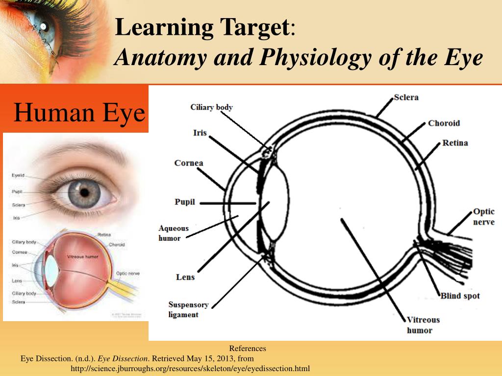
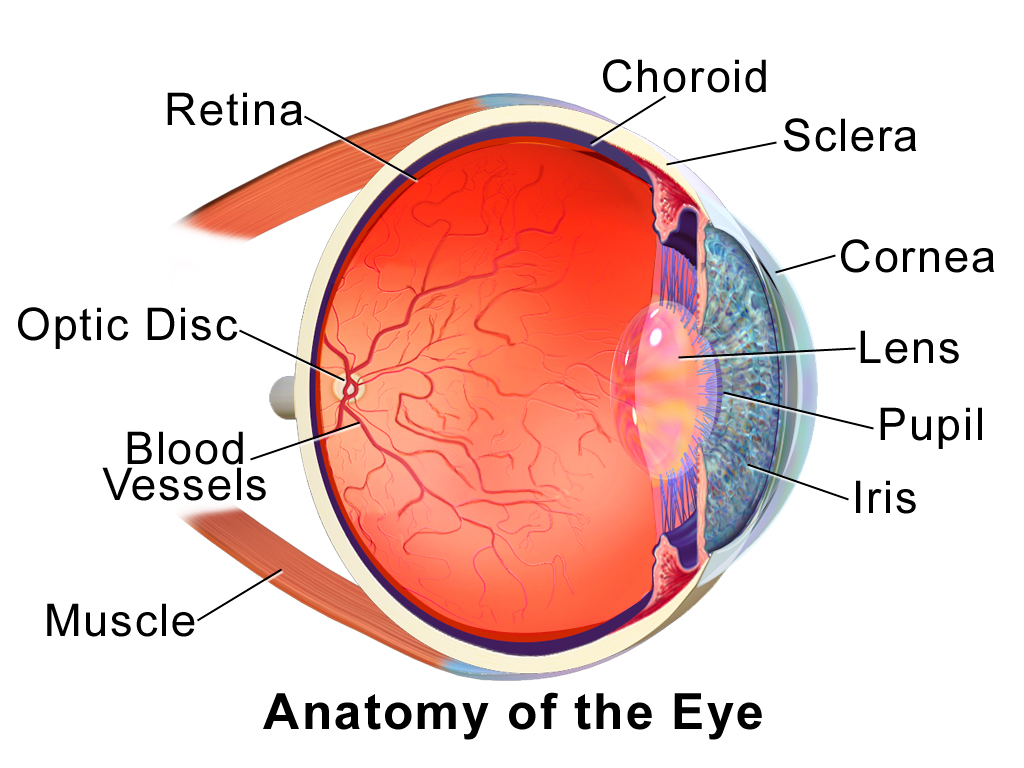
The sheep eye is approximately the same size as the human eye. It contains the same main ocular structures as of a human eye (see on diagram 1). This lab report of the sheep eye dissection will give a breakdown of all the different structures of a sheep eye. 1 pair of scissors
Sheep Eye Anatomy
Hold the eye so that the cornea is in an inferior position. 5. Making an incision into the eyeball about 1⁄2 cm from the edge of the cornea, cut completely around the eye (parallel to the cornea). Wear goggles to protect your eyes from the fluid that may spray out of the sheep eye. 6.

Sheep Eye Dissection YouTube
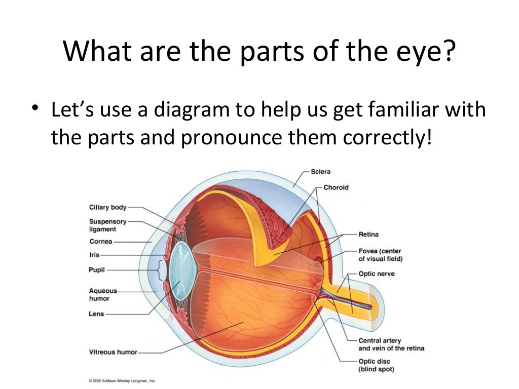
10. Choroid Layer, Tapetum lucidum Choroid Layer- lies between the sclera and the retina it provides the blood supply to the eye. Tapetum lucidum- iridescent film under the retina that provides animals with "night vision". Eye Dissection • Before we go over the dissection, let's review the parts of the eye and their function.
Sheep eye dissection
Sheep eye Dissecting pan Surgeon's gloves Scissors Single edge razor blade Probe Forceps Paper towels CONCEPTS: By dissecting and constructing labeled diagrams of eyes, students explore the structures and functions that contribute to the sense of vision.
Sheep Eye Dissection
Start studying Sheep Eye Anatomy. Learn vocabulary, terms, and more with flashcards, games, and other study tools.
Term 3 Sheep Eye Dissection Amanda's blog
Step 1: Wash the sheep eye in running water to remove the preservative fluid. Dry the eye with paper towel. Examine the front of the eye and locate the eye-lid, cornea, sclera (white of the eye) and fatty tissue. Examine the back of the eye and find extrinsic muscle bundles, fatty tissue and the optic nerve. The four extrinsic muscles (humans.